A New Look at Familiar Structures

As the innovation of medical practice continues to advance so does the need for novel teaching methods. Following the adoption of a new systems based, integrated medical curricula, the Department of Advanced Biomedical Education redoubled its efforts to bring new techniques into the preclinical classroom. These strategies included re-organizing the gross anatomy lab experience, expanding the virtual reality systems for gross anatomy, and the introduction of photogrammetric anatomical models.


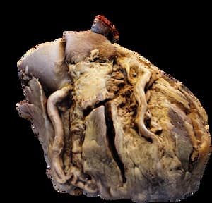
Photogrammetry is the process of converting 2D images of an object into 3D digital models. DABE faculty, in co-operation with the Office of Medical Education, have used these models to bring lab-like experiences into the traditional classroom setting. During the 2023-2024 academic year, M1 students were able to use 3D models of the donor hearts and kidneys they previously dissected during the Cardiovascular and Renal & Genitourinary System courses.
“Integrating photogrammetry into our curriculum has significantly enhanced student learning by providing an invaluable resource that is accessible from anywhere with internet access,” Dr. David Norris, Assistant Dean for Academic Affairs explains. “This continuity, using the very organs they dissected, reinforces their understanding and retention. Moreover, this approach supports adult learning principles and promotes active learning, engaging students in a dynamic and interactive educational experience.”
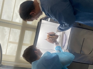
Photogrammetry also provides an opportunity to bring students into the process of educational research. Tyler Welch an M1 MSRP student and Aliecia Hyder a student in the Biomedical Masters program are working with Drs. Nathan Tullos and Erin Norcross to generate 3D models that will be used in the M2 preclinical curriculum.
Supporting the educational mission of the University by integrating basic science with clinical education is the primary goal of the Department of Advanced Biomedical Education. As Dr. Norma Ojeda, Chair of DABE explains, “Embracing visual technologies in medical training transforms abstract concepts into tangible experiences, enriching the integration of basic science with clinical practice.”
Medical education research continues to support the efficacy of active learning in pre-clinical education. Techniques and technologies such as virtual reality and photogrammetry can be used to ensure that the University continues to provide the best possible education for the future physicians of Mississippi.


